HUMAN MODELS OF DISEASE
Mechanistic insight is most informative when it can be tested in human systems that retain relevant genetic, developmental and physiological features. We build complementary hiPSC-derived models to determine how protein behaviour affects neurodevelopment, neurodegeneration and cardiac function, and to connect molecular mechanisms with tissue-specific human biology.
NEURODEVELOPMENTAL MODELS
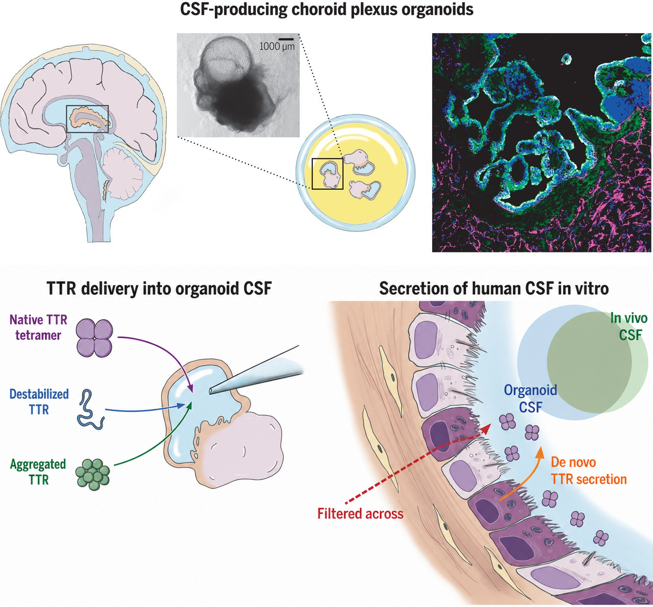
Human neural development depends on coordinated interactions among emerging neural tissues, extracellular signals and barrier-forming structures that cannot be fully reproduced in conventional monolayer cultures. We therefore develop organoid and engineered model systems that reproduce defined features of early human neural development while allowing experimental control over cellular composition and tissue interactions.
A central focus is the choroid plexus, a specialized structure that contributes to the environment in which the developing brain forms. We generate organoids that form choroid plexus structures alongside developing neural tissue and use these models to determine how the choroid plexus emerges, how it changes the developmental environment and how TTR influences neural development. Microfluidic platforms complement the organoids by controlling the spatial and temporal interaction of tissues, soluble factors and extracellular environments. Integrating these approaches distinguishes the physiological role of TTR during development from its later association with amyloid disease and provides experimental access to developmental relationships that are difficult to isolate in conventional culture.
HUMAN MODELS OF NEURODEGENERATION
Neurodegenerative disease emerges from interactions among genetic background, protein pathology and cellular stress that cannot be fully reproduced in traditional overexpression systems. Patient-derived hiPSC neurons preserve the genetic context in which pathology develops, while isogenic controls allow specific mutations or risk mechanisms to be examined against a matched background.
With a focus on Parkinson's Disease and related disorders, we use 2D cultures in combination with 3D organoid model systems to determine whether metabolic and mitochondrial deficits initiate neuronal pathology or amplify damage caused by disease-associated proteins. We then examine how these processes disrupt redox balance, mitochondrial quality control, cytoskeletal organization and the long-range axonal transport required to maintain neurons and synapses. By relating these changes to synaptic structure, calcium signalling and neuronal communication, we connect the biochemical state of aggregated proteins to early functional deficits that may precede neuronal loss. Collectively, these human models provide a mechanistic bridge from genetic and protein-level pathology to the selective dysfunction of vulnerable neurons.
Make it stand out
Human neural development depends on coordinated interactions among emerging neural tissues, extracellular signals and barrier-forming structures that cannot be fully reproduced in conventional monolayer cultures. We therefore develop organoid and engineered model systems that reproduce defined features of early human neural development while allowing experimental control over cellular composition and tissue interactions.
A central focus is the choroid plexus, a specialized structure that contributes to the environment in which the developing brain forms. We generate organoids that form choroid plexus structures alongside developing neural tissue and use these models to determine how the choroid plexus emerges, how it changes the developmental environment and how TTR influences neural development. Microfluidic platforms complement the organoids by controlling the spatial and temporal interaction of tissues, soluble factors and extracellular environments. Integrating these approaches distinguishes the physiological role of TTR during development from its later association with amyloid disease and provides experimental access to developmental relationships that are difficult to isolate in conventional culture.
HUMAN MODELS OF NEURODEGENERATION
Neurodegenerative disease emerges from interactions among genetic background, protein pathology and cellular stress that cannot be fully reproduced in traditional overexpression systems. Patient-derived hiPSC neurons preserve the genetic context in which pathology develops, while isogenic controls allow specific mutations or risk mechanisms to be examined against a matched background.
With a focus on Parkinson's Disease and related disorders, we use 2D cultures in combination with 3D organoid model systems to determine whether metabolic and mitochondrial deficits initiate neuronal pathology or amplify damage caused by disease-associated proteins. We then examine how these processes disrupt redox balance, mitochondrial quality control, cytoskeletal organization and the long-range axonal transport required to maintain neurons and synapses. By relating these changes to synaptic structure, calcium signalling and neuronal communication, we connect the biochemical state of aggregated proteins to early functional deficits that may precede neuronal loss. Collectively, these human models provide a mechanistic bridge from genetic and protein-level pathology to the selective dysfunction of vulnerable neurons.
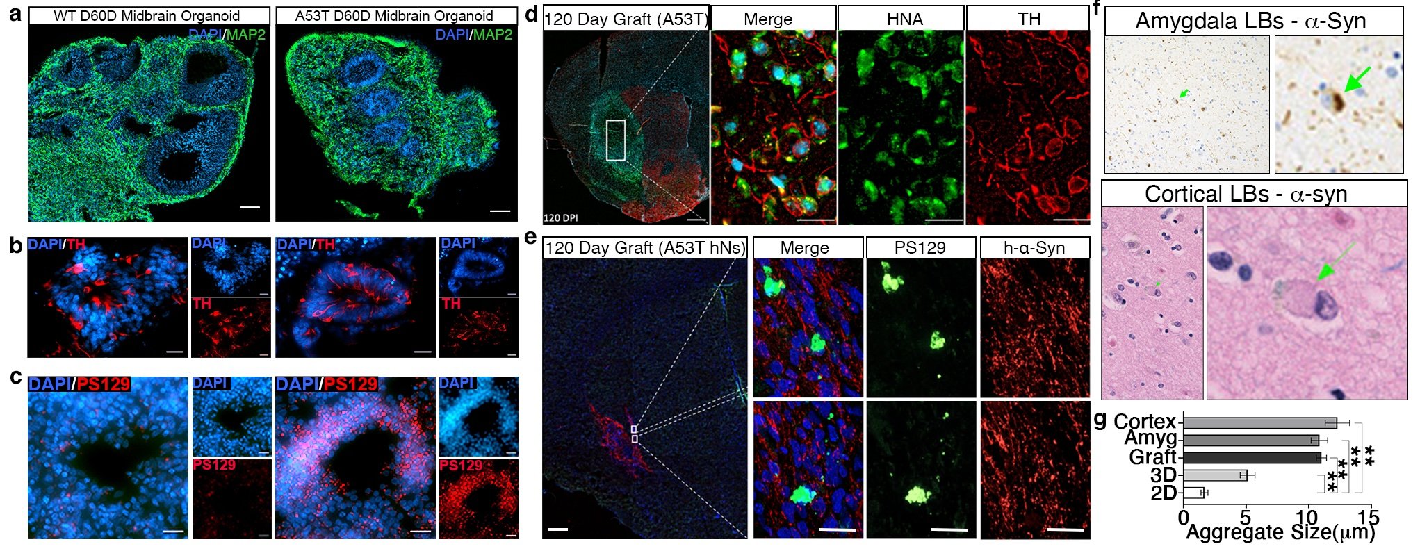
PD neurons exhibit greater pathology and larger aggregates when cultured for extended periods in either 3D organoids or following cell engraftment. a–c, Organoids derived from A53T and WT hESCs stained for MAP2 (a, neurons), TH (b, dopaminergic neurons) and pS129 α-synuclein (c). d–e, A53T hiPSCs were grafted into the putamen of 6-week-old NOD scid gamma (NSG) mice. At 120 days post-engraftment, sections were stained for human nuclear antigen (HNA) and TH (d). Antibodies specific for human α-synuclein (h-α-syn) were co-stained with pS129 (e). f, PD tissue immunolabeled for α-synuclein shows Lewy bodies in the amygdala and cortex. g, Organoid and engraftment models show aggregates similar in size to Lewy bodies.
HUMAN CARDIAC MODELS OF HEART FAILURE AND ATTR AMYLOIDOSIS
HUMAN CARDIAC MODELS OF HEART FAILURE AND ATTR AMYLOIDOSIS
Heart failure is a complex endpoint produced by convergent changes in metabolism, contractile function and tissue homeostasis. Transthyretin amyloid cardiomyopathy adds a distinct protein-assembly mechanism in which misfolded TTR accumulates within the heart and contributes to progressive dysfunction. Human induced pluripotent stem cell-derived cardiac models provide a defined system in which these mechanisms can be examined in a relevant human genetic and cellular context.
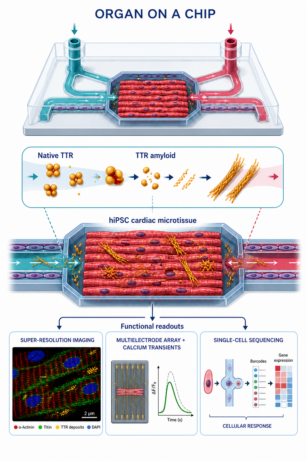
We develop hiPSC-derived cardiac models that reproduce defined cellular features of heart failure and allow us to determine how metabolic dysfunction, cellular stress and impaired contractile organization interact within human cardiomyocytes. In parallel, we investigate how TTR amyloid alters cardiomyocyte structure, metabolism and function. Integrating molecular and cellular measurements with functional assessment establishes when metabolic or protein-homeostasis deficits emerge and how they relate to impaired cardiac performance. Together, these models define how metabolic and amyloid mechanisms converge in heart failure and ATTR cardiomyopathy.
Make it stand out
Heart failure is a complex endpoint produced by convergent changes in metabolism, contractile function and tissue homeostasis. Transthyretin amyloid cardiomyopathy adds a distinct protein-assembly mechanism in which misfolded TTR accumulates within the heart and contributes to progressive dysfunction. Human induced pluripotent stem cell-derived cardiac models provide a defined system in which these mechanisms can be examined in a relevant human genetic and cellular context.
We develop hiPSC-derived cardiac models that reproduce defined cellular features of heart failure and allow us to determine how metabolic dysfunction, cellular stress and impaired contractile organization interact within human cardiomyocytes. In parallel, we investigate how TTR amyloid alters cardiomyocyte structure, metabolism and function. Integrating molecular and cellular measurements with functional assessment establishes when metabolic or protein-homeostasis deficits emerge and how they relate to impaired cardiac performance. Together, these models define how metabolic and amyloid mechanisms converge in heart failure and ATTR cardiomyopathy.